Waaaaaay back in grade school we learned of the double-helix structure of DNA. Ever since then, I’ve seen DNA represented in popular media in one of two ways. Lacking the ability to post pics here, I’ll merely link to the Wikipedia page about Watson and Crick’s landmark 1953 paper that first described that structure. That page itself shows two different representations of DNA:
First, in Figure 2. We see the double-helix that I’ve always visualized. Imagine a stepladder. Define an axis right up its middle, and then twist the ladder about that axis. Each rung is always perpendicular to (and bisected by) the twist axis.
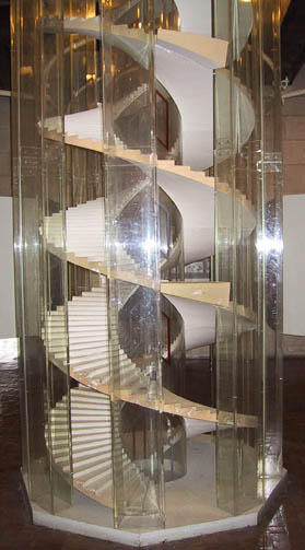
Second, in Figure 3. Imagine a stepladder. Lay it tangent to a cylinder whose diameter is maybe twice the width of the ladder. Set the ladder at some non-orthogonal angle so that when you wrap the ladder around the cylinder, it spirals up the cylinder’s length. The rungs (and legs) of the ladder lay flat against the surface of the cylinder.
Depending on which news program you’re watching, you might see one depiction or the other. So which one is correct?
I think one of them is just the result of the helix being tighter. I think if you were to take figure 3 and pull the ends a bit it would turn in to figure 2.
Here is a link of an article with an electron microscope image of DNA. Judge for yourself. I think it probably looks a bit more like the first one, but it’s hard to tell.
I know what you’re saying but that isn’t what double helix means. Figure 2 is used because showing DNA in a double helix is unnecessary to describing how it works.
You don’t have to draw a steam engine in 3D to describe its inner workings for instance.
I need to refresh my memory of the composition of DNA and bond angles. For now, I lean to the twisted stepladder.
I doubt DNA starts with the right angles and single plane of a stepladder. Carbon doesn’t bond like the, easy to draw on paper, right angles you see in books. Instead, it bonds with atoms in the corners of a tetrahedron. Only if double bonded is it planar, and those bonds are at 120 degrees. The amide linkage holding DNA together will have the 120 degree bonding in both the carboxyl radical and the amine.
All those rings and it has been so long I forget the names for the 2 stereoisomers rings come in or which is more common. Double bonds come in cis and trans, with cis dominating in nature, =/, trans /=/.
If you look at the animation for your cite you’ll notice that the axis the helix rotates on is crooked…as it must be otherwise the animation would be wobbly on it’s second axis.
Wiki is wrong, misunderstood, or misrepresenting the facts.
Neither figure is particularly accurate, although figure 3 is closer to the usual biologically-active form of DNA: B-DNA. Note the wider major groove and the narrower minor groove. Also commonly encountered are A-DNA, which has tighter coils, and the left-handed Z-DNA.
EDIT: And it’s called a double helix because there are two strands twisted around each other.
EDIT2: Biologists are not geometers. We can take forgive a little misalignment of our axes.
I’ll concede the double helix is called that because there are two edges, but I’m still absolutely convinced that the axis itself rotates around a second axis.
Are you suggesting that if you look at DNA from the top down, so that the perimeter that you see is mostly a circle, there would be no central “atrium” in DNA?* That is, that the points where the “spokes” from one level meet the one before and after don’t lie along a line,* but themselves spiral around a bit? I wonder that, too.
*Does any of this make sense? As those who’ve read my spinning top question know, I am no good at these sorts of descriptions.