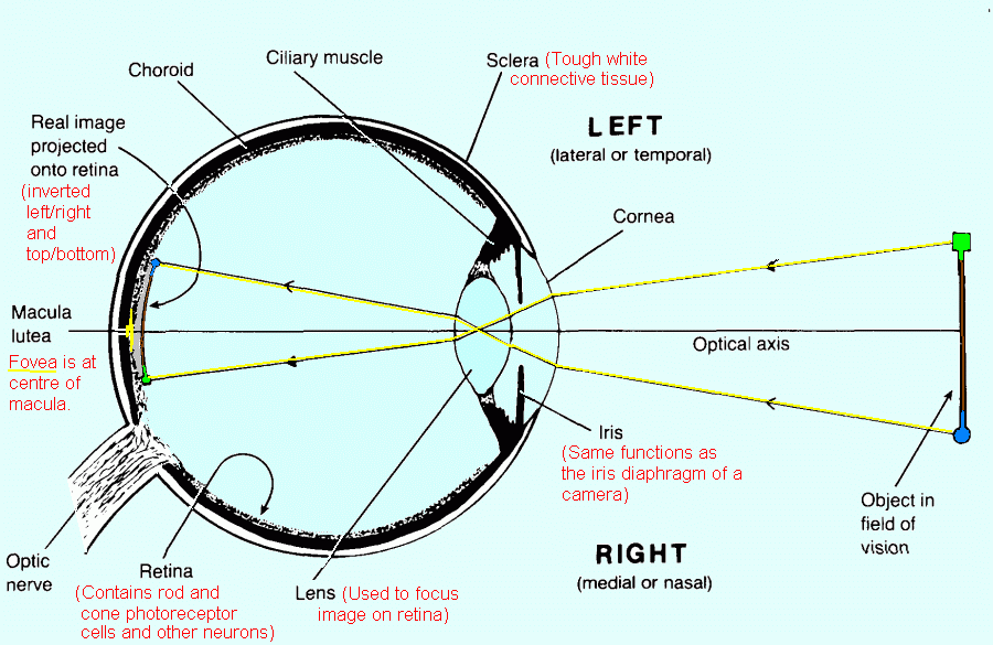
In school, I learned that being farsighted is caused by the lens in your eye being to weak and the focal point being behind the retina, while being near sighted is caused by the lens being to strong and the focal point being in front of the retina. So that would mean that in the nearsighted eye would have an inverted image hitting the retina, and the farsighted eye would have an upright image hitting the retina. So shouldn’t one of these situations result in upside down vision? The reason I asked about farsightedness is becuase I was also told the the image that usually hits the retina is inverted.
Whether nearsighted, perfectly sighted, or farsighted the image that hits the retina is always inverted - the question is whether it’s “in focus”.
Imagine manually focusing an old style film camera, the image does not flip from upright to inverted (or vice versa) when the focus is adjusted in front of or behind the film plane, it just gets blurry.
The image inversion actually occurs within the lens itself. The focal plane/point is different.
Not only that, but your brain would correct the image. What you see is what your brain interprets the image to be. They ran experiments with people wearing glasses that inverted the image to the eye. The brain corrected this after a while. When the glasses were removed the brain had to relearn not to correct the image. So I imagine even if your eye somehow always received an inverted image, you would never know it.
Supposing you were neither near- nor far-sighted. Is the image aligned correctly? Slightly shifting the focus in front of or behind the retina has no effect on it reversing top to bottom.
If I remember correctly, the image is always upside down, and the brain compensates for it. Like when you holod a magnifying glass at arms length.
Suppose you started off slightly nearsighted, and then gradually shifted to standard vision, and further to slightly farsighted. At what point would you expect the image to flip? Very slightly farsighted should be almost indistinguishable from normal or very slightly nearsighted.
What the OP is doing is confusing the rays in your linked image, which shows that rays of light from different objects or parts of an object end up on the “wrong” part of the retina, with those in this image Wikipedia:hyperopia which shows that different rays from the same point of an object are “inverted” when hitting the retina in far-sightedness.
I think it’s an understandable error to make, but as everyone has said it doesn’t matter for how the image is perceived. What’s happening is that light from the same exact point of the object that takes a lower path hits higher, and light on a higher path hits lower. But since it’s light from the same point, you get blurriness, not image inversion.
If you want to make this clearer, Kasavive, try drawing two extra lines alongside each of the two lines in cmyk’s picture. You should do it so the light from the top point of the object hits a large spot on the bottom of the retina, and the light from the top point hits a large spot on the top. Add the three lines for light from the central part of an object and it should be abundantly clear. ![]()
I think I know what the OP is getting at. The lens of the eye creates an inverted real image on the retina. The OP is probably imagining a convex lens diagram, where the image becomes virtual and upright when the object is moved outside the focal point.
One thing to keep in mind is that, even if this were a reasonable analogy, virtual images cannot be projected onto anything. If the light is not projected onto the retina, it is not seen. So this would not create an reverse image, regardless.
yes, it’s very annoying, I’ve had this misunderstanding, too. I’ve honestly seen two typical diagrams over and over again, the one where the focal point is in the middle of the eye and the image hitting the retina is inverted, and one where the focal point is on the retina?
As I posted earlier, the two typical diagrams show two different things. The behaviour of single lines of light from separate parts of an object, and the behaviour of separate lines of light from a single part of an object.
I’ve never seen one with the focal point in the middle and an inverted image. The images for hyperopia always show light from a single point, and no image.
I wasn’t going to post here, because I thought the first few posts covered it, but I see that there’s still misunderstanding here.
The diagram isn’t correct. The focal point is very close to the retina. Lemme explain.
In the first place, you should understand that, even though we have what is called a lens in the eye, which even looks like a glass lens, it’s not where most of the bending takes place. The reason for this is that you get the biggest effect where there’s a big index difference, and the “crystalline lens” in the eye is mostly saline embedded in the “vitreous humor”, which is also mostly saline. There’s not a lot of index change there. That lens is for fine tuning.
The big index change is at the cornea, where you’ve got air on one side (index = 1.0003) and mostly saline on the other ( n about 1.33). THAT’s where the bending takes place. Most of your optical power is right at the surface of the eye.
Next, what is the focal length? There are formulas for figuring out the location of the image , given the object distance and focal length, but they’re often given in confusing form. Here’s the simple form – don’t measure the distance of object or image from the lens. Measure it from the closest focal point*. And don’t measure it in feet, or inches, or millimeters. Measure the distances in units of the focal length.
now something interesting happens. The image distance measured from the focal point is the inverse of the object distance. What’s more, ythe magnification is just equal to the image distance (measured, recall, in units of the focal length).
If your object is one focal length from the focal point (which means it’s two focal lengths from the lens), then the image will be ne focal length from the other focal point, putting it also two focal lengths from the lens. It’ll also be at unit magnification (object and image are the same size, but upside-down relative to each other).
If the object is two focal lengths awaty, the image will be 1/2 of a focal length from the focal point.
If the object is three focal lengths, the image will be 1/3 of a focal length from the focal point.
If most of the objects are many focal lengths away, your images are all going to be very close to the focal point. That’s what happens in your eye. That image notwithstanding, your retina is just a short distance beyond the focal length. That way infinitely distant objects (stars, for instance), and far-away objects (that tree on that hill over there) and even only moderately distant objects (the blackboard, if you’re sitting in class) will all be pretty close to in-focus at the same time.
If something gets too close, or if you’re myopic (near sighted) or hyperoptic (far sighted), the image is out of focuys, but doesn’t flip upside-down. It just goes oput of focus.
I dealt with all of this in a slightly different form recently – whjat do thjings look like when you look through another lens? When does the image flip over? I had been told that this occurs when the lens is one focal length from the object you’re loking at. That seems to make sense, at first, but it’s wrong. An object viewed through a lens gets out of focus when you move it too close or too far from the lens, but it doesn’t “flip over” as you go beyond one focal length. It hits that point (where the image first fills the aperture – the image doesn’t abruptly “flip”) when the image produced by the lens sits exactly at the front focal point of the imaging system viewing it – your eye, or a camera. The lens will actually be more than a focal length away from the object when this happens.
*It’s not always the closest focal point, but it is in this circumstance.
I took that picture of the eyeball, and made three copies, showing the ray paths when the person is near-sighted, has good vision, and is far-sighted. This basically shows what others have been saying, but may make it clear for anyone who still doesn’t quite get it.
With good vision, any point in the field of vision focuses to a single point on the retina. For the near-sighted case, the rays focus in front of the retina, so they make a spot on the retina instead of a point, which makes vision blurry. For far-sighted vision, they focus behind it, but still make a spot, not a point.
For all three cases, the image is inverted, with the image of the green square below the image of the blue circle.
Nicely done. But, if the focal point isn’t on the fovea, the retina will not have the resolving power to distinguish between well focused and poorly focused points anyway. (And I rather doubt whether the eye even attempts to produce an optically sharp focus elsewhere but on the fovea.)
That’s great (I was tempted to do something similar!).
I think it helps demonstrate a focal plane (and a focal point on that plane) much better than words to.
