ssia.
Graphic pic here.
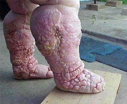
I’m guessing elephantiasis.
I thought the same thing, but does it cause pus and yellowness as seen in the picture? I would guess it does.
Leprosy?
I’m siding with Guin and dig. When you have an incredible amount of lymph fluid that cannot be drained from the tissue I would think that all sorts of pus (from battling infections) and other necrosis to the tissue would be possible.
Here’s an elephantiasis photo that looks markedly different:
That first picture looks like a skin disorder…ultra psoriasis or something.
There are some skin diseases like scleroderm that cause thickening of the skin. Doesn’t look like elephantiasis to me. I’ll see if I can pull a photo and pop back in.
Nope. Here’s scleroderma. Not ichthyosis, either.
Elephantiasis is often complicated by fungal and bacterial infections of the skin.
That’s the condition that severly obese people sometimes get, right? Because that’s what I was thinking it mgiht be, since I didn’t think elephantitis got all scabby and pussy like that.
But, of course, lymphedema doesn’t seem to get too scabby, either. And leprosy doesn’t cause that kind of swelling.
[intercom]
Dr.Mercotan to the GQ ward, Dr.Mercotan to the GQ ward.
[/intercom]
Neurofibromatosis?
Chromomycosis?
This looks to me like the late-stage skin changes of chronic lymphedema, with inflammatory fibrosis of the skin and subcutaneous tissues. Recurrent, chronic bacterial and/or fungal infections can lead to some very deforming skin and soft tissue changes. Soft, ‘pitting’ edema is gradually transformed into woody, granular scar tissue.
As far as a definitive diagnosis goes, I don’t think that you’ll get one, because the skin changes seen here can be the end result of any number of initial insults - radiation, lymph node injury, hereditary diseases, or vascular abnormalities - or even combinations of things like heart failure and diabetes, or obesity and venous thrombosis.
Most of the scleroderma patients that I have seen have thinning and tightening of the skin, rather than thickening like this. Elephantiasis can result in this sort of damage, which is why it is managed in part by careful attention to skin hygeine so as to prevent recurrent skin infections and stave off this kind of scarring.
Might I recommend purulent? I know we all say it, but remember how it looks in the chart  .
.
One thing’s for damn sure, though - this person shouldn’t be walking around outside barefoot! Wrap those rascals!
Might I ask where the OP found the picture?
I’m not an MD, and I’ve never seen elephantiasis. However, something about the flamingo-pink color and the very clean skin texture points to Hollywood special effects, in my opinion. Somebody who could keep it that clean could probably have access to good medical care, and it never would have gotten that bad.
Throwing my hat into the ring…Myxedema
This was a biopsy proven case and I took the picture myself.
THe photo in the OP could also by plain old lymphedema. To add to the confusion, a form of lymphedema closely resembling the photo in the OP is termed elephantiasis verrucosa nostra.
-Choosy Derm MD
Although I’m sure that I could be wrong and all these answers look good, I would really like to hear some explanations about why they *do not * think this is elephantiasis. I know the skin looks more infected and necrotic than a lot of the “classic” photos of the disease, but I don’t understand how that rules it out.
It could be an old, old photo of someone with Guinea Worm. It has been extinct for some time now.
I am guessing from the number of lesions in the upper level of the skin. Or something else.